What is Retroperitoneum Organs?
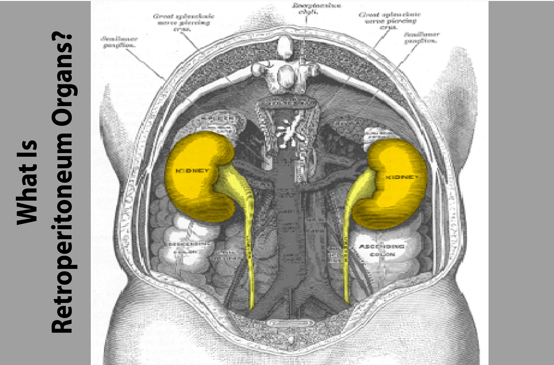
Retroperitoneum Organs mesothelioma, also called retroperitoneal peritoneal mesothelioma, is the name for cancer that affects the peritoneal organs, or organs located in the abdomen. The peritoneum is the covering of the organs that lies between the rectum and the diaphragm. These organs are made up of several organs that are needed for proper digestion and other normal metabolic functions. The peritoneum consists of the colon, the duodenum, the small intestines, the large intestines, the liver, spleen, pancreas, and some glandular tissue as well. Organs that are directly attached to the visceral wall, or peritoneum, are retroperitoneal organs in the abdomen.
Types of Retroperitoneum Organs
There are five primary retroperitoneal organs, which are the spleen, the intestine, the liver, the diaphragm, and the upper part of the small bowel. These organs are located in the lower part of the abdominal cavity, or duodenum. The first two are located inside the rectum while the last two are located outside the duodenum. This may seem confusing because the organs actually share similar traits with each other, such as the location, but they are actually quite different in function and anatomy. The small intestine is the tube-like organ that empties into the abdomen while the liver is an active part of the body that produces bile.
Two Major Roles Retroperitoneum Organs
The retroperitoneum, however, has two major roles. The first role is that of a protective sac that covers the peritoneum and prevents fluid from entering the abdominal viscera. The second role is that of an immune system that produces antibodies that attack cells that are outside of the abdominal viscera. These retroperitoneal antibodies often cause inflammation or scarring of the intestinal lining.
Causes of Retroperitoneum Organs
One of the more common causes of inflammation of the retroperitoneum is the presence of interstitial fluid filled with cytokines. Which are generally secreted by the white blood cells. This interstitial fluid serves to protect the retroperitoneum. But in some cases, it can also promote the growth of both cancerous and non-cancerous cells. Cancerous cells can frequently form tumors in the retroperitoneum. While non-cancerous cells can frequently form adhesions or scar tissues around the organs in the duodenum and liver. These adhesions can restrict the movement of the organs and reduce their functionality.
Peritoneal Mesotheliomas
Peritoneal mesotheliomas are the most common retroperitoneal cancer type. They generally begin with the transformation of a normal cell (leukemia) into a cancer cell (leptomeningitis). Leukemia is also a leading cause of cirrhosis and dialysis in the general adult population. As a result, the majority of persons afflicted with leukemia will suffer from serious abdominal pain, polyps, and peritoneal masses. These structures usually take the form of either a solid mass or an ulcerated soft lump. Leukemia is frequently characterized by large numbers of immature blood cells (granulocytic cells) or by multiple, diffuse scars.
Abdominal Cavity
The retroperitoneum covers approximately half of the abdominal cavity. So the majority of the structures within this area are directly exposed to cellular inflammation. A wide variety of cell and non-cellular debris accumulates here. The incidence of the inflammatory disease depends on the cellular debris and the thickness of the abdominal wall. This tissue is particularly thick in the upper GI tract and in the peritoneal cavity below the dextrocardia. In recent years, the parietal layers have been found to play a significant role in determining. The severity of abdominal pain in patients with peptic ulcers.
Parietal Cells
The parietal cells in the stomach are responsible for the attachment of the retrosternal organs to the stomach wall and for the localization and location of the portal vein, which drains the stomach fluids and the small intestine. The jejunum and the small intestine are connected to the colon via the transitional zone. The primary difference between the stomach and colon is that the colon has a significantly deeper layer of undigested material that lies just beneath the parietal cells. This material includes both the endocrine and gastrointestinal glands. The portal vein drains off the liver into the small intestine. The portal vein also drains into the right lower colon (the jejunum) and into the right upper GI tract (the jejunum and ileum).
Also Read: Schmorls Nodes Root Disorder
The Retroperitoneal Structures May Divide Into Two Types
The papillary type, which has only a single kidney (sometimes referred to as the aplys), and the multivesicular type, which has two or more kidney organs, although not necessarily in a conformation that is typical of their respective organs. Of the two types, the multivesicular is the most common, being present in over 90% of the population. The remaining less common types have either a single kidney or an additional kidney. The retroperitoneal glandular structures are situated close to the adrenal glands. Where the pituitary gland synthesizes cortisol, the primary stress hormone.