What is Phrenic Nerve?
The term “phrenia” refers to a group of approximately thirty different species of sensory nerves or afferents. That is distributed in various ways throughout the entire nervous system. Most people have two sets of sensory nerves. The median nerve in the head and the median nerve in the neck; the median nerve exits through the top of the head and passes through the heart to exit the lungs; the neck nerve exits through the neck and goes through the lungs. The other twelve nerves are distributed in various ways. One set exits via the spinal cord to the base of the skull, another sets down the back of the tongue, and a fourth goes to the ankle. Each set of twenty to thirty sensori-nostril fibers has a common extension along the posterior spine and extends up to about one inch inside the body cavity.
Branches of Phrenic Nerve
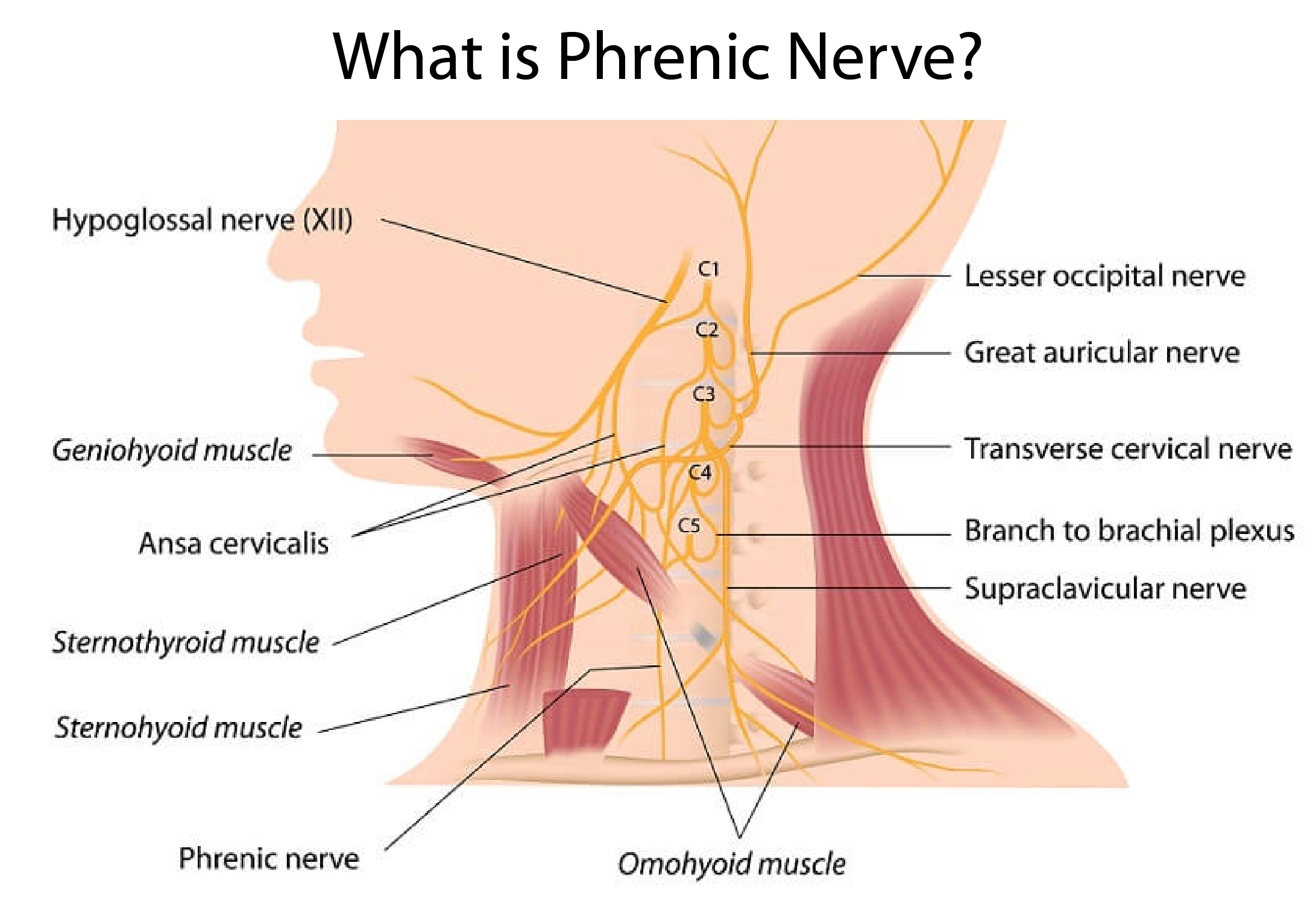
The phrenic nerve usually originates in the middle to lower part of the neck through the C2 curve to the diaphragm and passes through the heart and lungs. From its source, the phrenic nerve branches over to the upper chest and then down the right side of the body to the chest wall. In rare instances, the branch extends to the stomach.
First Part of Phrenic Nerve
The first part of the nervous system is the accessory nervous system (also called the peri-allergic division). This division includes the eyes, nose, mouth, limbs, and cervical spine. In the neck, the accessory phrenic nerve controls the movement of the external auditory canal, which is important for hearing. In the superior cervical region, the superior epigallocate nerves control breathing through the upper respiratory tract and provide the sensory inputs to the eye for sight.
When Phrenic Nerve Becomes Injured
If the accessory phrenic nerve becomes injured, this division controls difficulty breathing. This difficulty breathing creates a medical condition known as hypoxemia. As a result of this disorder, the patient may experience breathlessness even when the heart is pumping blood into the body. Sometimes, even if the injured nerve is not actually directly pinched to the diaphragm muscle, the sensation of breathlessness will be triggered when the injured nerve has been compressed to an area close to or exactly within the pericardium muscle.
When this happens, the injured nerve can no longer deliver pain signals to the brain because it has become pinched between the diaphragm and the pericardium. Thus, it becomes possible for the hemidiaphragm to move to relieve this pressure. This movement of the hemidiaphragm triggers contractions in the chest wall muscles (the Pectoralis minor and Pectoralis major). Which expands the chest cavity to facilitate the flow of air into the lungs and out of the lungs. This opening of the chest cavity facilitates a rise in cardiac output. The exact mechanisms underlying the action of this reflex are not clearly understood but are believed to involve muscular activation of the heart.
Coronary Artery
The action of the diaphragms and the coronary artery during respiration causes variation in the blood pressure that reaches the brain. The location and size of the brain’s internal pacemaker. Which is located inside the skull. Determine where this blood flow will go. The action of the Pectoralis minor and Pectoralis major muscles is particularly important. Because these two muscles help control the direction of this ventricular flow. The ventricular rate (VAT) is determined by the rate of blood flow to and from the heart and is thus a key determinant of brain blood pressure. The location of the superior vena cava and paraventricular valve (the spongy tissue within the belly of the heart). Also plays a role in the functioning of the brain’s blood vessels.
Imbalance of Blood Pressure
An example of a common phrenic nerve disorder is carpal tunnel syndrome which develops. When a bulging or inflamed posterior chest wall causes compression of the inferior vena cava. Which then creates an “imbalance” of blood pressure in the neck. Phosphate. A normal function of the anterior chest wall muscles becomes “stuck” in the narrowed opening of the trunk. When the muscles are contracted. This causes pressure to be exerted on the spinal cord. The result is a pain in the neck.
Lumbar Subluxation
A second common cause of phrenic nerve stimulation is the development of bilateral diaphragmatic paralysis or CDP. Cervical Spondylosis is the development of an abnormally shaped spinal column that impairs its normal function. It has been found that Cervical Spondylosis can result from many different causes including trauma, tumor, or infection, or can even be the result of congenital abnormalities. A typical case involves the diagnosis of spondylolisthesis or lumbar subluxation caused by an accident or by poor posture while carrying out specific activities of daily living.