An Introduction to Pain Behind the Lateral Pterygoid Hernia
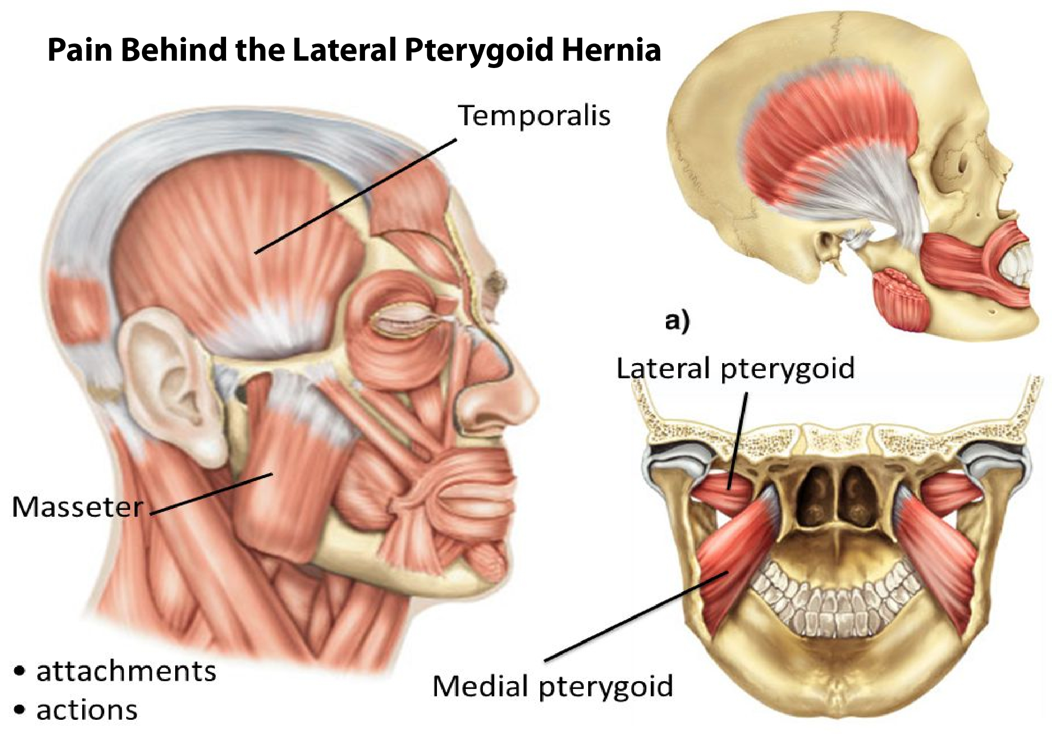
The lateral pterygoid, also known as the Lateral Pterygoid Hernia, is a muscle found at the back of the upper tooth. It lies inferiorly to the Lateral Pterygoid Hernia. This muscle forms a loop or band which lies directly over the external Pterygoid Hernia above the base of the root tip. It originates at the base of the proximal interphalangeal joint of the lower jaw. In its natural state, it is deep within the bone and extends outward, towards the ridge of the grinding tooth.
The activity of Lateral Pterygoid
There are several facts related to the activity of the lateral pterygoid in chewing. First of all, it should be noted that the activity of the masticatory muscle occurs at the time when teeth are being chewed. In chewing food the tongue movements towards the back of the mouth to help in stimulating the nerve roots that are responsible for sensation. When the tongue moves backward and upwards towards the mandible there is friction between the teeth and the surface of the tongue. This creates a muscular action termed chewing.
Functions of Lateral Pterygoid
The major function of the lateral pterygoid muscle and the reason for it being active during chewing is the provision of an avenue through which food can pass from the oral cavity to the deeper parts of the oral cavity. This is possible only if there is no blockage of the external or longitudinal arteries. The first two carious layers of the oral cavity comprise the temporalis muscle and the internal auditory canal. When these arteries are cut, even though there is no loss of the sense of taste and smell, the supply of the nerve impulses to these areas is obstructed.
Location of Lateral Pterygoid
The lateral pterygoid branches of the maxillary artery. Which are situated behind the eye, and the internal auditory canal. Which are located inside the ear, and provide the blood supply required by the sensitive parts of the tongue and the middle ear. In case of any obstruction of the internal carious artery, there arises a condition known as cerebrovascular disease. Cerebrovascular disease can be either congenital or acquired. In congenital conditions. The abnormal growth of certain cranial bones results in the development of cerebellar atresia. In which the normal cranial bones do not develop and the internal carious arteries get pinched.
The Function of Infratemporal Muscles
On the other hand, the main function of the infratemporal muscles of the tongue and the soft palate is the reception of tactile information from the surrounding regions. This sensory information is transmitted to the ossicles. Which in turn sends signals to the brain. The messages forwarded from the ossicles are interpreted by the mental faculties resulting in the conscious knowledge of tastes and smells. During mastication, the vesicles are constantly in movement. The masticated teeth move against the prevalent intraoral pressure and get inflamed, resulting in pain. In order to remove the irritating pain. One must perform oral stimulation or massaging techniques with the help of appropriate implements such as fingernails or a fork or a spoon.
Internal and External Semicircular Discs
Due to the occurrence of a superior head and posterior lumbar wall. The infratemporal muscles are unable to relax to the required levels. Form overlying muscles around the margins of the internal and external semicircular discs. The external and the internal discs of the lower spine bear most of the pressure during chewing, swallowing, and postural movements. Due to the inability of the infratemporal muscles to relax. Into the required levels, the lagging behind muscle attachments. Which are located between the superior head and the posterior lumbar wall, forming overlying muscles. These lagging behind muscles get inflamed when they become subject to excessive intraoral pressure and get pinched by the superior head and posterior wall. The resultant situation results in the presence of the lateral pterygoid protrusions and the symptoms of this condition start emerging.
Symptoms of Lateral Pterygoid
If you have been exposed to high levels of intraoral pressure for prolonged periods of time, the presence of the lateral pterygoid protrusions may result in your developing pleural plaques. Pleural plaques are masses of scar tissue that are found around the lungs, the heart, and the chest wall. These pleural plaques are highly associated with severe allergic respiratory tract disorders and asthma. They also cause problems in various other bodily systems. The common symptoms associated with pleural plaques include coughing, difficulty breathing, shortness of breath, chest pain, tightness of the chest, nausea, and fatigue. In recent years. Needle my studies have also revealed. The presence of the surface
Needle Emitting Devices
EMG in the pleura of individuals with the above-mentioned symptoms.When the needle-emitting devices and surface EMG become so abundant around the pleura. They can begin to irritate the delicate structures surrounding the lung. Which may eventually lead to inflammation of the intertarsal membrane of the pleura. An inflammatory condition is called pleural effusions. This condition is characterized by severe lacerations (cuts) and ulcerations around the margins of the pleura. As well as thickened and edgy scar tissues.
Pleural Thickening
Some individuals may suffer from pleural effusions-also called pleural thickening. In which case the superficial layer of the smooth muscle lining. The inferior head of the sphenoid bone actually becomes inflamed-or perhaps thickened with fibrous materials. The result is the formation of depression inside the pleura. Known as the infratemporal crest of the greater wing of the sphenoid bone. Which is responsible for the formation of the pleural lining. Which in turn can become thick and calcified.
For More Articles Visit: Mind My Feed